Importance of evaluating the uterine cavity before in vitro fertilization
Evaluating the uterine cavity before in vitro fertilization is a crucial step in the assisted reproduction process. In this blog, you’ll discover the importance of performing this evaluation and the different methods used, such as hysteroscopy and hysterosonography. You will also learn how we use hysterosonography at Panama Fertility and the advantages it offers over vaginal ultrasound, achieving success rates of up to 97%.
What is in vitro fertilization and why is it important to evaluate the uterine cavity?
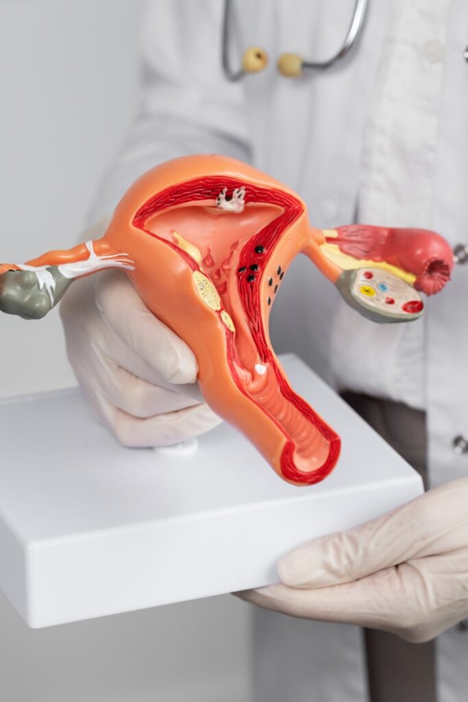
In vitro fertilization or ivf is an assisted reproduction procedure in which eggs are fertilized outside the woman’s body, in a laboratory. It is an option for couples who have difficulty conceiving naturally. Before undergoing this procedure, it is crucial to evaluate the uterine cavity to ensure it is an optimal environment for embryo implantation. A healthy uterine cavity increases the chances of success of in vitro fertilization and reduces the risk of complications.
Evaluating the uterine cavity allows for the detection of possible anomalies or problems that could affect embryo implantation, such as polyps, fibroids, adhesions, or uterine malformations. Identifying these issues before the ivf procedure allows for the necessary measures to be taken to correct or treat them, thus improving the chances of treatment success.
Evaluating the uterine cavity allows for the detection of possible anomalies or problems that could affect embryo implantation, such as polyps, fibroids, adhesions, or uterine malformations. Identifying these issues before the ivf procedure allows for the necessary measures to be taken to correct or treat them, thus improving the chances of treatment success.
Methods of evaluating the uterine cavity
There are several methods used to evaluate the uterine cavity before in vitro fertilization. Two of the most common methods are hysteroscopy and hysterosonography (saline infused sonogram).
Hysteroscopy is a procedure in which a hysteroscope, a thin tube with a light and a camera at the end, is introduced through the vagina and cervix into the uterus. This allows the doctor to directly examine the inside of the uterine cavity and detect any anomalies. During hysteroscopy, biopsies or therapeutic procedures can also be performed, if necessary.
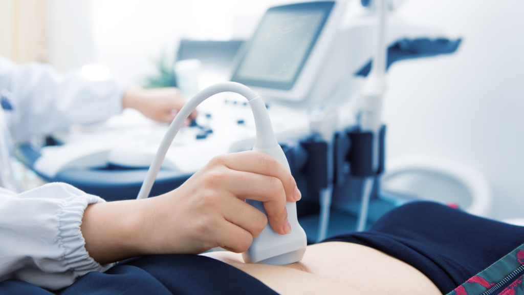
Hysterosonography, also known as saline infusion sonography, is an advanced technique that combines ultrasound and the infusion of saline solution into the uterine cavity. It is used to assess the shape and size of the uterus, as well as to detect polyps, fibroids, or other anomalies. During hysterosonography, saline solution is introduced into the uterus through a catheter, allowing for better visualization of the uterine cavity on ultrasound.
These evaluation methods provide detailed images of the uterine cavity and detect any abnormalities that could affect the success of in vitro fertilization.
Hysteroscopy is a procedure in which a hysteroscope, a thin tube with a light and a camera at the end, is introduced through the vagina and cervix into the uterus. This allows the doctor to directly examine the inside of the uterine cavity and detect any anomalies. During hysteroscopy, biopsies or therapeutic procedures can also be performed, if necessary.
Hysterosonography, also known as saline infusion sonography, is an advanced technique that combines ultrasound and the infusion of saline solution into the uterine cavity. It is used to assess the shape and size of the uterus, as well as to detect polyps, fibroids, or other anomalies. During hysterosonography, saline solution is introduced into the uterus through a catheter, allowing for better visualization of the uterine cavity on ultrasound.
These evaluation methods provide detailed images of the uterine cavity and detect any abnormalities that could affect the success of in vitro fertilization.
Hysteroscopy: a precise and safe method
Hysteroscopy is considered a precise and safe method for evaluating the uterine cavity before in vitro fertilization. It allows for direct visualization of the uterine cavity and the possibility of performing biopsies or therapeutic procedures if necessary. This procedure is performed on an outpatient basis and generally does not require general anesthesia, making it a comfortable and safe option for patients.
During hysteroscopy, a hysteroscope is used that is introduced through the vagina and cervix into the uterus. The doctor can examine the uterine cavity and perform any necessary intervention in the same procedure. Hysteroscopy allows for a precise and direct evaluation of the uterine cavity, making it a reliable method for detecting possible abnormalities that could affect in vitro fertilization.
During hysteroscopy, a hysteroscope is used that is introduced through the vagina and cervix into the uterus. The doctor can examine the uterine cavity and perform any necessary intervention in the same procedure. Hysteroscopy allows for a precise and direct evaluation of the uterine cavity, making it a reliable method for detecting possible abnormalities that could affect in vitro fertilization.
Hysterosonography: an advanced technique for evaluating the uterine cavity
Hysterosonography, also known as ultrasound with saline infusion, is an advanced technique used to evaluate the uterine cavity. It combines ultrasound with the infusion of saline solution into the uterus, allowing for better visualization of the uterine cavity and the detection of possible abnormalities.
During hysterosonography, a catheter is introduced into the uterus through the vagina and saline solution is infused. The saline solution expands in the uterine cavity, providing greater clarity in ultrasound images. This allows the doctor to identify polyps, fibroids, or other anomalies that could affect in vitro fertilization. Hysterosonography is a safe technique and well tolerated by patients, providing accurate information about the uterine cavity.
At Panama Fertility, we use hysterosonography as part of our evaluation of the uterine cavity before in vitro fertilization. This advanced technique allows us to obtain detailed and precise images of the uterine cavity, helping us to identify any issues that could affect the success of the treatment. Hysterosonography has proven to be a useful tool at Panama Fertility, as it has allowed us to achieve success rates of up to 97% in our in vitro fertilization treatments.
Advantages of hysterosonography over vaginal ultrasound
Hysterosonography offers several advantages over vaginal ultrasound in evaluating the uterine cavity before in vitro fertilization.
First, hysterosonography allows for better visualization of the uterine cavity. The infusion of saline solution into the uterus expands the cavity, facilitating the detection of possible abnormalities, such as polyps or fibroids. In comparison, vaginal ultrasound may not provide as clear an image of the uterine cavity.
Moreover, hysterosonography is a less invasive technique than vaginal ultrasound. While vaginal ultrasound requires the insertion of a probe into the vagina, hysterosonography only requires the introduction of a catheter into the uterus. This makes it more comfortable and tolerable for patients.
Another advantage of hysterosonography is that it allows for a more accurate evaluation of the uterine cavity. The infusion of saline solution improves the quality of ultrasound images, making it easier to detect any anomaly.
In summary, hysterosonography offers significant advantages over vaginal ultrasound in the evaluation of the uterine cavity before in vitro fertilization. It is a more precise, less invasive technique and provides better visualization of the uterine cavity.
These advantages have contributed to the high level of success we have achieved at Panama Fertility using hysterosonography in our in vitro fertilization treatments.
Conclusions
Evaluating the uterine cavity before in vitro fertilization is essential to increase the chances of success of the treatment and reduce the risk of complications. Both hysteroscopy and hysterosonography are effective and safe methods for evaluating the uterine cavity.
Hysteroscopy allows for direct visualization of the uterine cavity and the performance of therapeutic procedures if necessary. Hysterosonography, on the other hand, combines ultrasound with the infusion of saline solution to obtain detailed images of the uterine cavity.
At Panama Fertility, we use hysterosonography as part of our evaluation of the uterine cavity before in vitro fertilization. This advanced technique has allowed us to achieve success rates of up to 97% in our treatments.
In conclusion, evaluating the uterine cavity before ivf using methods such as hysteroscopy and hysterosonography is crucial for achieving successful outcomes in assisted reproduction treatments. If you have had previous failed assisted reproduction treatments, it is important to perform an adequate evaluation of the uterine cavity.